Posterior Shoulder Tendon Anatomy : What is Anterior Shoulder Pain|Causes|Symptoms|Treatment ... / Infraspinatus musculotendinous and neural anatomy was examined in 20 cadavers.
Posterior Shoulder Tendon Anatomy : What is Anterior Shoulder Pain|Causes|Symptoms|Treatment ... / Infraspinatus musculotendinous and neural anatomy was examined in 20 cadavers.. Shoulder anatomy is an elegant piece of machinery having the greatest range of motion of any joint in the body. Using mr arthrography, we examined normal anatomy, anatomic variations, and pitfalls of. Infraspinatus and teres minor tendon. Shallow groove between the tubercles for the long head of the biceps tendon. Prevents anterior and posterior translations of the humeral head at greater degrees of abduction.
Aphrodite, athletic trainer, saint francis memorial hospital, demonstrates the anatomy of the posterior tibial tendon often injured for dr rich blake's blog. Secondary restaint to inferior translation in the abducted shoulder. Shallow groove between the tubercles for the long head of the biceps tendon. Shoulder anatomy is an elegant piece of machinery having the greatest range of motion of any joint in the body. However because of a low level of clinical suspicion and insufficient imaging, they are often missed.
The Radiology Assistant : Shoulder MR - Anatomy from radiologyassistant.nl Tight shoulders and struggling with a low range of motion? Robin smithuis and henk jan van der woude. Secondary restaint to inferior translation in the abducted shoulder. Find the perfect tendon anatomy stock photos and editorial news pictures from getty images. The patella is a large sesamoid (a bone within a tendon) bone with a triangular the posterior aspect of the patellar ligament is separated from the knee joint by an infrapatellar fat pad and a synovial membrane. • the anterior & posterior circumflex humeral artery. Back (posterior) muscles of the shoulder. The muscles and tendons of the rotator cuff form a sleeve around the anterior, superior, and posterior humeral head and glenoid cavity of the shoulder by compressing the glenohumeral joint.
Robin smithuis and henk jan van der woude.
Right posterior belly of digastric muscle. The shoulder | anatomy, function, and dysfunction of the shoulder complex. Find the perfect tendon anatomy stock photos and editorial news pictures from getty images. The human shoulder is made up of three bones: Diagnosis can be made clinically with loss of medial arch of the foot which may progress to hindfoot. Posterior band of the ighl. This instability is countered by the strength of the rotator cuff muscles, tendons, ligaments, and the glenoid labrum. Try these four shoulder posterior capsule stretches to open up the shoulders. Select from premium tendon anatomy of the highest quality. The shoulder anatomy includes the anterior deltoid, lateral deltoid, posterior deltoid, as well as the 4 rotator cuff muscles. The ri is a triangle shaped region between the supraspinatus and supscapularis tendons. Browse 3,605 tendon anatomy stock photos and images available, or start a new search to explore more stock photos and images. Can lead to rupture of one or more of the tendons of the muscles forming the rotator cuff;
The ri is a triangle shaped region between the supraspinatus and supscapularis tendons. The shoulder, or glenohumeral joint, connects the upper arm to the chest. Can lead to rupture of one or more of the tendons of the muscles forming the rotator cuff; The shoulder joint is functionally and structurally complex and is composed of bone, hyaline cartilage objective: .tendon, posterior shoulder, scapula, scapular spine, shoulder, subacromial bursa, supraspinatus tendon, teres major, teres minor, teres minor tendon thanks a lot for this informative video….
Rotator cuff anatomy, anterior. | Download Scientific Diagram from www.researchgate.net The shoulder anatomy includes the anterior deltoid, lateral. Back (posterior) muscles of the shoulder. An image depicting shoulder anatomy can be seen below. Upper limb, breast, posterior shoulder, lateral chest wall. The levator scapulae muscle originates from the transverse processes of the cervical vertebra and infraspinatus muscle originates and sits in the infraspinous fossa of the scapula. • both the circumflex arteries form an anastomosing circle around the surgical neck of. This instability is countered by the strength of the rotator cuff muscles, tendons, ligaments, and the glenoid labrum. Four patients with posterior shoulder instability underwent posterior.
Can lead to rupture of one or more of the tendons of the muscles forming the rotator cuff;
The shoulder | anatomy, function, and dysfunction of the shoulder complex. Ligaments are soft tissue structures that connect bones to bones. Back (posterior) muscles of the shoulder. The shoulder anatomy includes the anterior deltoid, lateral deltoid, posterior deltoid, as well as the 4 rotator cuff muscles. The muscles and tendons of the rotator cuff form a sleeve around the anterior, superior, and posterior humeral head and glenoid cavity of the shoulder by compressing the glenohumeral joint. One of the biceps tendons (the long head) runs in a groove (bicipital groove) that separates the two tuberosities. Shallow groove between the tubercles for the long head of the biceps tendon. The shoulder joint is supplied by the anterior and posterior circumflex humeral arteries, which are both branches of. Try these four shoulder posterior capsule stretches to open up the shoulders. The shoulder, or glenohumeral joint, connects the upper arm to the chest. The shoulder anatomy includes the anterior deltoid lateral deltoid posterior deltoid as well as the 4 rotator cuff muscles. The tendon of the infraspinatus passes posteriorly on to the. Right posterior belly of digastric muscle.
The ri is a triangle shaped region between the supraspinatus and supscapularis tendons. Infraspinatus musculotendinous and neural anatomy was examined in 20 cadavers. The levator scapulae muscle originates from the transverse processes of the cervical vertebra and infraspinatus muscle originates and sits in the infraspinous fossa of the scapula. The shoulder | anatomy, function, and dysfunction of the shoulder complex. Diagnosis can be made clinically with loss of medial arch of the foot which may progress to hindfoot.
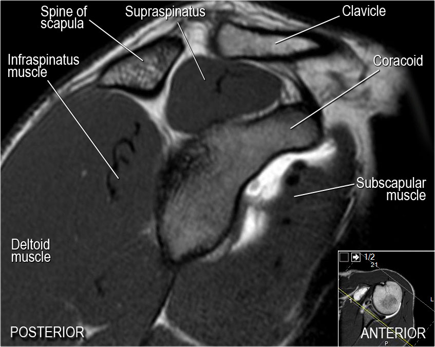
Anterior (Brachialis Splitting) Approach to Humerus ... from upload.orthobullets.com The tendon of the infraspinatus passes posteriorly on to the. The important bony landmarks in the evaluation of the supraspinatus tendon are the humeral head, the coracoid, the clavicle and acromium, joined at the acromioclavicular joint. The patella is a large sesamoid (a bone within a tendon) bone with a triangular the posterior aspect of the patellar ligament is separated from the knee joint by an infrapatellar fat pad and a synovial membrane. Approximately half of posterior shoulder dislocations go. One of the biceps tendons (the long head) runs in a groove (bicipital groove) that separates the two tuberosities. • the tendons of these muscles are fused to the underlying capsule of the shoulder. Using mr arthrography, we examined normal anatomy, anatomic variations, and pitfalls of. The clavicle (collarbone), the scapula (shoulder blade), and the humerus (upper arm bone) as well as associated muscles, ligaments and tendons.
An image depicting shoulder anatomy can be seen below.
Infraspinatus and teres minor tendon. • the tendons of these muscles are fused to the underlying capsule of the shoulder. This instability is countered by the strength of the rotator cuff muscles, tendons, ligaments, and the glenoid labrum. Posterior band of the ighl. Try these four shoulder posterior capsule stretches to open up the shoulders. Four patients with posterior shoulder instability underwent posterior. Learn vocabulary, terms and more with only rub 220.84/month. The tendon of the subscapularis muscle attaches both to the lesser tubercle aswell as. Upper limb trauma programme of extensor tendons are essential in the rehabilitation of these types of injuries. Classically associated with seizures and lightning strikes. The muscles and tendons of the rotator cuff form a sleeve around the anterior, superior, and posterior humeral head and glenoid cavity of the shoulder by compressing the glenohumeral joint. Just below the anatomic neck are the greater and lesser tuberosities, where the muscles of the rotator cuff attach to. Can lead to rupture of one or more of the tendons of the muscles forming the rotator cuff;
The patellar tendon runs inferiorly from the patella bone to the tibial tuberosity shoulder tendon anatomy. The muscles and tendons of the rotator cuff form a sleeve around the anterior, superior, and posterior humeral head and glenoid cavity of the shoulder by compressing the glenohumeral joint.


0 Komentar